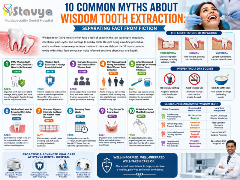
Wisdom teeth, anatomically known as the third molars, are the final set of permanent teeth to erupt in the human mouth. Typically emerging during the late teens or early twenties—a developmental period historically referred to as the “age of wisdom”—these four molars sit at the very back of the upper and lower dental arches.
While a small percentage of the population experiences properly aligned, fully erupted third molars that function harmoniously, the vast majority of modern humans face structural challenges. Due to evolutionary changes in dietary habits, the human jawbone has gradually evolved to be smaller over millennia, often leaving insufficient structural space to accommodate these late-emerging teeth.
When a wisdom tooth lacks the space to erupt normally, it becomes impacted—meaning it remains completely or partially trapped beneath the gum tissue or jawbone. Left untreated, an impacted or poorly positioned wisdom tooth can trigger a cascade of localized infections, severe nerve pain, structural damage to adjacent healthy teeth, and even bone-destroying cysts.
At Stavya Dental Hospital, our oral surgery and preventative dentistry teams prioritize evidence-based, conservative clinical care. Recognizing the early indicators of wisdom tooth complications can save you from debilitating dental emergencies and permanent structural damage. This comprehensive guide breaks down the science of third molar pathology, details the seven critical signs that mandate immediate surgical extraction, and outlines what to expect during a modern, pain-free extraction procedure.
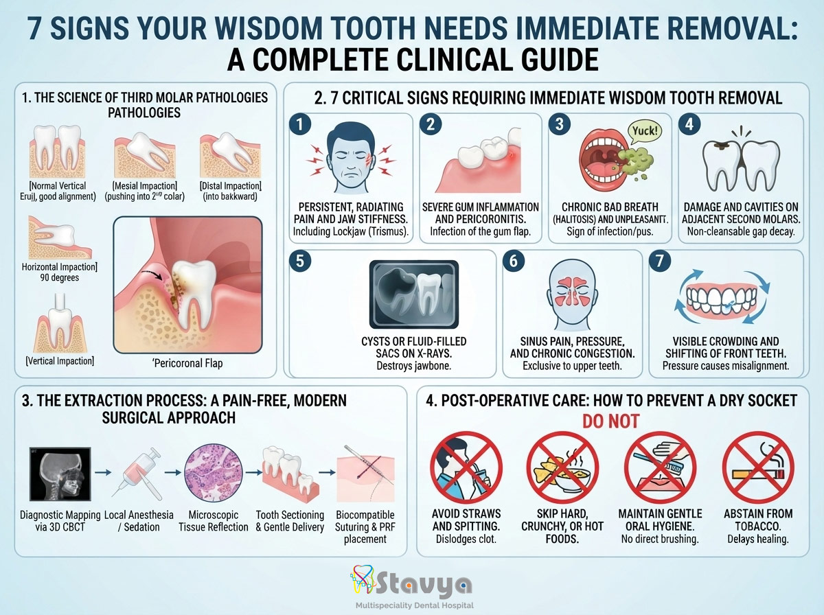
1. The Science of Third Molar Pathologies: Why They Fail to Erupt
To understand why wisdom teeth routinely require clinical intervention, it helps to look at how they develop. Because they are the last teeth to form, they must navigate a crowded oral environment. If the jawbone lacks adequate length, the wisdom tooth will attempt to erupt at abnormal, pathological angles.
[Normal Vertical Eruption]  --> Harmonious alignment (Rare)
[Mesial Impaction]          --> Tooth tilts forward, pushing directly into the 2nd Molar
[Distal Impaction]          --> Tooth tilts backward, burying into the rear jawbone
[Horizontal Impaction]      --> Tooth grows completely flat at a 90-degree angle
[Vertical Impaction]        --> Tooth is upright but trapped entirely beneath the bone
As a wisdom tooth struggles against structural barriers, it typically succumbs to one of several types of impaction:
- Mesial Impaction: The tooth tilts forward at an angle, directing its chewing surface straight into the root structure of the healthy second molar ahead of it.
- Horizontal Impaction: The tooth grows completely sideways at a sharp 90-degree angle. This orientation exerts continuous, aggressive mechanical pressure against the adjacent tooth roots and can cause severe bone loss.
- Vertical Impaction: The tooth is oriented in a normal upright position but remains entirely locked beneath the dense bone of the jaw, unable to break through the surface.
- Distal Impaction: The tooth tilts backward toward the rear of the jaw, getting trapped in the ascending ramus of the mandible.
When a tooth is partially impacted—meaning only a small portion of the crown breaks through the gum tissue—it creates a highly dangerous anatomical trap called a pericoronal flap. This loose flap of gum tissue acts as a microscopic pocket that easily traps food particles, oral bacteria, and plaque. Because it is physically impossible to clean beneath this flap with a standard toothbrush or dental floss, the area becomes an ideal breeding ground for aggressive anaerobic infections.
2. 7 Critical Signs Requiring Immediate Wisdom Tooth Removal
If you or a family member experiences any of the following symptoms, it indicates that the third molar has transitioned from a silent issue into an active clinical threat requiring immediate evaluation and surgical extraction.
Sign 1: Persistent, Radiating Pain and Jaw Stiffness
One of the most immediate indicators of a wisdom tooth complication is a dull, throbbing pain originating deep in the back of the jaw. This pain rarely stays localized; because the third molars sit close to major facial nerve pathways (such as the inferior alveolar nerve), the pressure from an impaction can cause radiating pain that travels upward into your ear, temple, throat, or neck.
As the inflammation spreads into the primary muscles responsible for chewing (the masseter and temporalis muscles), patients frequently develop a clinical condition called trismus—commonly known as lockjaw. This severely limits your ability to open your mouth comfortably, making eating, speaking, and basic oral hygiene highly painful.
Sign 2: Severe Gum Inflammation and Pericoronitis
When bacteria become trapped beneath the gum tissue surrounding a partially erupted wisdom tooth, it triggers an acute localized infection known as pericoronitis. The gum tissue in the very back of the mouth will appear deep red, visibly swollen, and highly tender to the touch.
As the infection progresses, even the gentle mechanical pressure of your upper teeth biting down can bruise the swollen tissue, intensifying the pain. In severe, unmanaged cases of pericoronitis, the infection can breach the localized tissues and spread deep into the spaces of the neck and face, presenting a systemic health risk that requires urgent clinical care.
Sign 3: Chronic Bad Breath (Halitosis) and an Unpleasant Taste
If you notice a foul, metallic taste in your mouth or chronic bad breath that persists despite meticulous brushing, tongue scraping, and mouthwash, the culprit is often a hidden wisdom tooth infection.
The inaccessible pockets around an impacted molar allow food particles to ferment and putrefy over time. This process fuels the growth of volatile sulfur compound-producing bacteria. Additionally, chronic inflammation often leads to the subtle, continuous drainage of pus from beneath the gum flap. This discharge causes a bitter or sour taste that signifies an active, deep-seated bacterial infection.
Sign 4: Damage and Cavities on Adjacent Second Molars
An improperly positioned wisdom tooth doesn’t just destroy itself; it actively threatens the health of the entire dental arch. When a third molar tilts forward and rests heavily against the adjacent second molar, it creates a tight, highly restrictive space known as a non-cleansable gap.
Plaque and food debris accumulate continuously within this junction. Because no toothbrush, interdental brush, or strand of floss can reach this area, aggressive decay develops simultaneously on the back of the healthy second molar and the front of the wisdom tooth. By the time a patient feels pain from this specific interaction, the decay has often penetrated deep into the nerve chamber of the second molar, requiring complex root canal therapy or, in worst-case scenarios, the extraction of both teeth.
Sign 5: Cysts or Fluid-Filled Sacs on Diagnostic X-Rays
Every tooth develops inside a protective, fluid-filled sac within the jawbone. Once a normal tooth erupts, this sac naturally dissipates. However, if an impacted wisdom tooth remains locked deep inside the bone for an extended period, the residual sac can fill with fluid, developing into a benign but destructive structure called a dentigerous cyst.
As the cyst slowly expands over months or years, it exerts outward hydraulic pressure that hollows out the surrounding healthy jawbone. This structural bone loss weakens the jaw, making it substantially more susceptible to fractures under minor trauma. Left completely unmanaged, these cysts can destroy localized nerve pathways and even transform into complex, aggressive tumors that require extensive bone grafting and reconstructive surgery.
Sign 6: Sinus Pain, Pressure, and Chronic Congestion
This specific symptom applies exclusively to the upper (maxillary) wisdom teeth. The roots of the upper third molars develop in close structural proximity to the floor of the maxillary sinuses—the air-filled cavities located behind your cheekbones and above your upper teeth.
As the roots of an upper wisdom tooth grow and push upward against the sinus floor, they can perforate or heavily irritate the delicate sinus membranes. This pressure manifests as chronic sinus congestion, unexplained headaches, a constant feeling of pressure beneath the eyes, and nasal drainage that does not respond to conventional allergy or cold medications.
Sign 7: Visible Crowding and Shifting of Front Teeth
While the orthodontic community continues to study the exact mechanical force an erupting wisdom tooth exerts on the entire dental arch, extensive clinical observation confirms that the continuous pressure from horizontally or mesially impacted molars can cause gradual tooth movement.
As the third molars push forward to find a path out of the bone, they can trigger a domino effect across the dental arch. This pressure can cause previously straight teeth—including teeth carefully aligned through years of orthodontic braces—to slowly rotate, overlap, and crowd forward. Extracting the wisdom teeth early neutralizes this force, preserving the structural integrity and alignment of your smile.
3. The Extraction Process: A Pain-Free, Modern Surgical Approach
A primary reason many individuals delay essential wisdom tooth care is dental anxiety or a fear of surgical pain. Fortunately, modern oral surgery has advanced dramatically, transforming wisdom tooth extraction into a highly predictable, comfortable, and completely pain-free outpatient procedure.
Phase 1: Comprehensive Digital Diagnostics
At Stavya Dental Hospital, we never operate blindly. The extraction journey begins with high-resolution digital imaging, utilizing Panorex X-rays or advanced 3D Cone Beam Computed Tomography (CBCT) scans. This technology lets our oral surgeons map out the exact three-dimensional position of the tooth roots relative to major anatomical structures, such as the inferior alveolar nerve canal in the lower jaw and the sinus cavities in the upper jaw, ensuring maximum surgical safety.
[Diagnostic Mapping via 3D CBCT]
       |
[Administration of Local Anesthesia / Sedation] --> Complete numbness achieved
       |
[Microscopic Tissue Reflection] --> Minimizes trauma to surrounding gums
       |
[Tooth Sectioning & Gentle Delivery] --> Prevents heavy mechanical stress on jawbone
       |
[Biocompatible Suturing & Platelet-Rich Fibrin (PRF) placement] --> Accelerates healing
Phase 2: Comfort and Anesthesia Administration
Before any surgical step begins, the area is completely numbed using advanced local anesthetics. For patients experiencing severe dental anxiety or those requiring the simultaneous removal of all four impacted molars, modern sedation options are available. This ensures you remain completely relaxed, comfortable, and entirely unaware of the surgical mechanics throughout the appointment.
Phase 3: Conservative Surgical Technique
If a wisdom tooth is buried beneath the bone, the oral surgeon makes a small, precise incision in the gum tissue to gently expose the tooth structure. Rather than pulling the tooth out in a single piece—which can apply unnecessary force to the surrounding bone—modern surgeons use a technique called tooth sectioning. The molar is safely divided into smaller pieces, which are delivered one by one through a conservative opening. This approach preserves the surrounding bone and significantly reduces post-operative swelling and discomfort.
4. Post-Operative Care: How to Prevent a Dry Socket
Your recovery experience depends heavily on the care habits you practice during the first 48 hours following surgery. Proper self-care protects the surgical site and accelerates your body’s natural healing mechanisms.
Understanding and Preventing a Dry Socket (Alveolar Osteitis)
When a tooth is removed, your body naturally forms a stable blood clot inside the empty bone socket. This clot acts as a protective blanket, shielding the exposed bone and sensitive nerve endings underneath while new tissue grows over them.
If this blood clot is accidentally dislodged, dissolved, or washed away prematurely, it results in a highly painful condition called a dry socket. This exposes the raw bone directly to air, fluids, and food debris, causing a sharp, radiating pain that typically begins 3 to 5 days after surgery.
To protect the blood clot and ensure a smooth recovery, adhere to these essential guidelines:
- Avoid Straws and Spitting: The negative pressure created inside your mouth when sucking through a straw or spitting forcefully can cleanly dislodge the blood clot from its socket.
- Skip Hard, Crunchy, or Hot Foods: Stick strictly to a soft, cool diet for the first few days (e.g., yogurt, smoothies, lukewarm blended soups, mashed potatoes). Hot liquids can dissolve the delicate clot, while sharp foods like chips can physically puncture the healing tissue.
- Maintain Gentle Oral Hygiene: Do not brush directly over the surgical site for the first 24 hours. Afterward, rinse your mouth very gently with warm salt water after meals instead of rinsing aggressively or spitting.
- Abstain from Tobacco: Smoking or using chewing tobacco introduces toxic chemicals that delay tissue repair, while the physical act of inhaling severely increases your risk of developing a dry socket.
5. Prioritizing Your Oral Health at Stavya Dental Hospital
At Stavya Dental Hospital, we approach oral surgery with a deep focus on patient comfort, safety, and long-term wellness. Our surgical suites are equipped with advanced diagnostic tools, including ultra-low-dose 3D CBCT imaging, which allows our specialists to plan and execute complex third molar extractions with micro-precision.
We emphasize a transparent, supportive approach to patient care. From your initial diagnostic consultation through your final follow-up checkup, our team walks with you every step of the way, providing detailed, tailored recovery steps and personal care support. By addressing impacted or symptomatic wisdom teeth before they cause widespread infection or damage neighboring teeth, we help you protect your long-term oral health and maintain a comfortable, pain-free smile.
Schedule Your Advanced 3D Wisdom Tooth Consultation Today
If you or a family member is experiencing jaw pain, swelling, or changes in your bite, do not wait for a major dental emergency to develop. Contact our expert team today to schedule an advanced diagnostic evaluation.
- Official Website: www.stavyadental.com
- Direct Clinic Helpline: +91 8980395039