Root canal therapy (endodontic treatment) has a remarkable track record. It is one of the most effective dental procedures available, boasting success rates between 85% and 95% when performed by an experienced clinician. The primary objective is straightforward: eliminate deep-seated infection from inside a tooth’s root canal system, seal the space to keep bacteria out, and save the natural tooth from extraction.
However, like any complex medical or dental intervention, a root canal can occasionally experience complications. A tooth that underwent successful treatment months or even years ago can suddenly begin to ache, develop sensitivity, or show signs of a renewed infection on a routine dental X-ray.
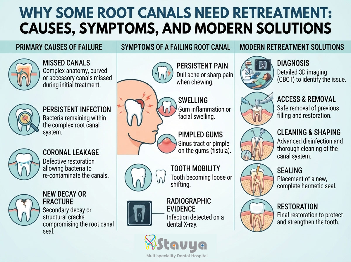
When an initial endodontic treatment fails to heal or becomes reinfected, root canal retreatment is often the best line of defense to preserve the tooth.
At Stavya Dental Hospital, we emphasize clinical transparency and patient education. In alignment with modern digital health communication guidelines, this comprehensive, evidence-based guide explores the anatomical and clinical reasons why some root canals require retreatment, how to recognize the symptoms of a failing root canal, and what to expect during the corrective procedure.
1. The Anatomy of a Root Canal: Why It’s a Complex Procedure
To understand why a root canal can fail, it helps to look closely at the internal anatomy of a human tooth.
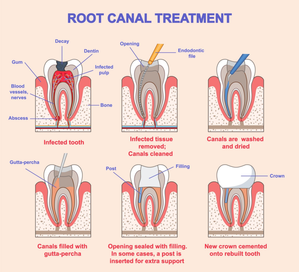
The Complex Internal Anatomy and Standard Steps of Root Canal Therapy.
A tooth is not a solid block of enamel. Beneath the hard outer enamel and dentin layers lies a hollow chamber containing the dental pulp—a delicate network of nerves, blood vessels, and connective tissues. This pulp extends from the center of the crown down through the roots via narrow pathways called root canals.
While textbooks often depict root canals as straight, uniform tubes, real human teeth are vastly more complex. A single root canal can feature:
- Microscopic Lateral Canals: Tiny branch lines branching off the main canal like roots of a tree.
- Severe Curvatures: S-shaped or sharply hooked roots that make physical access difficult.
- Canal Bifurcations: A single canal that splits into two separate channels near the tip (apex) of the root.
- C-Shaped Configurations: Complex web-like canal networks commonly found in lower second molars.
If even a microscopic pocket of bacteria or necrotic (dead) tissue is left behind in one of these intricate web-like spaces, an infection can slowly rebuild over time.
2. Primary Reasons an Initial Root Canal Fails
When a root canal fails, it rarely means the initial dentist made a mistake. More often, it is a reflection of the tooth’s unique biological challenges or events that occurred long after the initial procedure was completed. Clinically, these factors are categorized into immediate procedural challenges and long-term delayed complications.
Immediate and Structural Causes
1. Narrow or Curved Canals Overlooked During the First Procedure
Some root canals are so microscopic or calcified (hardened by age or chronic trauma) that they are entirely invisible to the naked eye. If a tooth has four root canals but only three are located, cleaned, and sealed, the tissue left inside the hidden fourth canal will eventually break down, harbor anaerobic bacteria, and cause a recurrent infection at the base of the root.
2. Delayed Placement of the Permanent Restoration (Crown)
Once the internal root canal system is cleaned and sealed with a biocompatible material called gutta-percha, the tooth is vulnerable. The temporary filling placed at the end of the appointment is only designed to last a few weeks. If a patient delays returning to the clinic for a permanent dental crown or definitive filling, the temporary seal will eventually degrade under the force of chewing. Saliva, which is highly rich in oral bacteria, will leak inside the tooth, recontaminating the root canals.
3. Inadequate Seal or Microleakage
Even if a permanent crown is placed promptly, it must achieve a perfect, hermetic marginal fit against the natural tooth structure. If there is a microscopic gap along the edge of the crown, a process called microleakage occurs. Over months or years, saliva and bacteria slowly seep underneath the restoration, travel past the dental cement, and find their way down into the root fillings.
Delayed Causes (Years After the Procedure)
1. New Tooth Decay (Secondary Caries)
A tooth that has had a root canal can no longer feel pain from hot, cold, or sweet triggers because its nerves were removed. However, the outer structure of the tooth is still vulnerable to tooth decay. If oral hygiene slips, a new cavity can form at the margin where the crown meets the natural tooth structure. If this decay left untreated, it will burrow deep into the center of the tooth, causing a secondary infection of the root canal system.
2. Structural Fractures or Cracks
Teeth that have undergone root canal therapy become inherently more brittle over time because they no longer have an internal blood supply to keep the dentin hydrated. If the tooth is subjected to extreme biting forces—such as chronic teeth grinding (bruxism) or biting down hard on an unpopped popcorn kernel—it can develop an invisible fracture. A vertical root fracture that travels down the root allows bacteria from the gums to flood directly into the bone surrounding the tooth, causing a fast-moving, painful infection.
3. Symptoms of a Failing Root Canal: When to Seek Treatment
A failing root canal doesn’t always hurt immediately. In fact, many secondary infections are completely asymptomatic in their early stages and are only discovered during routine dental checkups. However, when symptoms do emerge, they serve as warning signs that the surrounding jawbone is under threat.
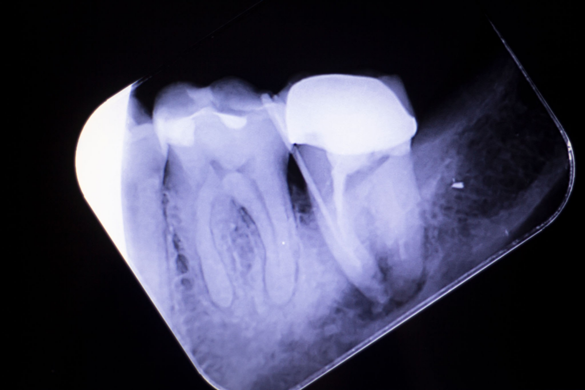
An X-Ray View of an Endodontic Lesion (Dark Shadow) at the Root Tip.
If you notice any of the following symptoms, it is crucial to seek a professional evaluation:
- Persistent Dull Ache or Throbbing Pain: A continuous, deep ache inside the jawbone or localized to the specific tooth, which may worsen when lying down or during changes in physical posture.
- Pain When Chewing or Applying Pressure: Since the internal nerve of the tooth is gone, pain upon biting down indicates that the infection has spread out of the tooth and into the surrounding periodontal ligament and jawbone, making the bone socket inflamed and tender.
- Swelling of the Gums Near the Affected Tooth: The gum tissue near the root tip may look red, swollen, or feel tender to the touch.
- A “Pimple” on the Gums (Sinus Tract): A persistent bump on the gums, known clinically as a fistula or sinus tract, may form. This acts as a drainage channel for pus generated by a bone-level abscess. It may periodically drain a bitter-tasting fluid, providing temporary relief from pressure and pain.
- Mobile or Loose Tooth: As an untreated infection destroys the supporting alveolar bone around the roots, the tooth may begin to shift or feel loose in its socket.
4. Root Canal Retreatment vs. Tooth Extraction: Making the Right Choice
When faced with a failing root canal, patients often ask, “Why shouldn’t I just pull the tooth and be done with it?” While extracting a problematic tooth provides immediate relief from symptoms, it introduces a series of long-term structural and financial challenges that make saving the natural tooth via retreatment a far superior choice in most clinical scenarios.
Why Saving the Natural Tooth is Preferred
No prosthetic replacement—no matter how advanced—can completely match the biological performance of a natural tooth. Your natural tooth roots are wrapped in a highly specialized ligament that acts as a natural shock absorber when you chew and sends precise sensory feedback to your brain.
Furthermore, keeping your natural tooth keeps your smile aligned. When a tooth is extracted, the neighboring teeth lose their lateral support and begin to tilt and drift into the open space, disrupting your bite and potentially leading to temporomandibular joint (TMJ) disorders.
Comparing Your Long-Term Clinical Options
| Clinical Consideration | Endodontic Retreatment | Tooth Extraction & Dental Implant |
| Primary Objective | Save and disinfect the natural tooth structure. | Remove the tooth and replace it with a titanium screw. |
| Invasiveness | Minimally invasive; works entirely inside the existing tooth. | Requires surgical extraction and bone healing phases. |
| Treatment Timeline | Typically completed in 1 to 2 clinical visits. | Can take 3 to 6 months for bone integration before final placement. |
| Bone Preservation | Preserves the natural bone surrounding the root. | Requires an implant or bone graft to prevent bone resorption. |
| Long-Term Success | Very high if root anatomy can be fully accessed and cleaned. | High, but requires healthy bone density and systemic health. |
While a dental implant is an excellent solution when a tooth is genuinely unsavable (such as in the case of a severe vertical root fracture), root canal retreatment remains the most conservative, cost-effective, and fast-acting method to restore your oral health.
5. What to Expect During a Root Canal Retreatment Procedure
The thought of undergoing a second root canal on the same tooth can feel daunting. However, thanks to modern dental anesthetics and advanced micro-endodontic technologies, the retreatment experience is highly predictable and virtually identical to a standard deep filling appointment in terms of comfort.
The clinical procedure follows a strict, highly controlled protocol designed to identify and eliminate the hidden causes of the initial failure:
Step 1: Comprehensive Diagnostic Imaging
Before any treatment begins, high-resolution diagnostic imaging is performed. At Stavya Dental Hospital, we utilize advanced CBCT (Cone Beam Computed Tomography) 3D Scans. Unlike a flat, traditional 2D X-ray, a CBCT scan provides a 3D rendering of your tooth and jawbone. This allows our specialists to view the tooth from every angle, locate curved or hidden canals, and pinpoint the exact boundary of the bone infection before making an opening.
Step 2: Gaining Access to the Root Canals
The tooth is completely numbed using localized anesthetics to ensure a pain-free experience. A protective rubber sheet called a dental dam is placed over the tooth to isolate it from saliva and bacteria. If the tooth has a dental crown, a small, precise opening is created through the top of the crown to gain access to the inside of the tooth.
Step 3: Removal of Existing Filling Materials
The specialist carefully removes the old gutta-percha filling material and any posts or core materials that were placed inside the root canals during the first procedure. This is done using specialized ultrasonic instruments that gently loosen the material without damaging the surrounding natural dentin structure.
Step 4: Advanced Disinfection and Shaping
Once the canals are empty, the interior of the tooth is thoroughly inspected under high-magnification endodontic microscopes. The canals are thoroughly flushed with chemical disinfecting solutions to kill any lingering bacteria, and flexible nickel-titanium rotary instruments are used to reshape the canals, ensuring every microscopic corner is completely clean.
Step 5: Resealing and Restoring the Tooth
After verifying that the canals are completely sterile and dry, they are packed with a fresh layer of gutta-percha to form a long-lasting, hermetic seal against future bacterial invasion. A temporary filling is placed over the opening, and after a short healing phase, a new, custom-fit permanent crown is placed to fully restore the tooth’s structural strength and appearance.
6. Your Partners in Endodontic Health: Stavya Dental Hospital
At Stavya Dental Hospital, we treat root canal retreatment not just as a routine repair job, but as an advanced, precision-guided clinical intervention. We combine specialized clinical training with state-of-the-art diagnostic and therapeutic technologies to maximize the chance of saving your tooth, even in highly complex or challenging cases.
If you are experiencing pain, swelling, or sensitivity in a tooth that has previously had a root canal, early diagnostic intervention is the key to avoiding an extraction.
Schedule an Advanced Endodontic Evaluation
Protect your natural smile and resolve chronic dental discomfort today. Contact our specialist clinic to book a comprehensive diagnostic consultation.
- Official Website: www.stavyadental.com
- Direct Contact Helpline: +91 8980395039