Modern dentistry relies heavily on what cannot be seen by the naked eye. While a clinical visual examination is the foundation of any dental checkup, more than half of each tooth structure—along with the surrounding jawbone, nerve pathways, and sinus cavities—lies hidden beneath the gum tissue. To diagnose disease, plan complex surgeries, or evaluate structural growth, dental professionals must rely on advanced diagnostic imaging.
For decades, traditional two-dimensional (2D) dental X-rays were the gold standard for looking beneath the surface. However, the introduction of Cone Beam Computed Tomography (CBCT) has sparked a technological revolution in dental diagnostics. By shifting from flat 2D images to fully interactive three-dimensional (3D) digital models, CBCT has fundamentally changed how dentists diagnose oral pathologies and plan treatments.
At Stavya Dental Hospital, our clinical philosophy centers on diagnostic precision, patient safety, and evidence-based treatment mapping. Patients frequently ask us: What is the difference between a CBCT scan and a traditional X-ray, and which one is better for my dental care?
This comprehensive guide breaks down the science behind both imaging systems, compares their clinical advantages, and explains how advanced 3D diagnostics ensure safer, more predictable outcomes for your smile.
1. The Physics and Mechanics: How Dental Imaging Works
To understand the practical clinical differences between these two technologies, it helps to look at the underlying engineering of how they capture data.
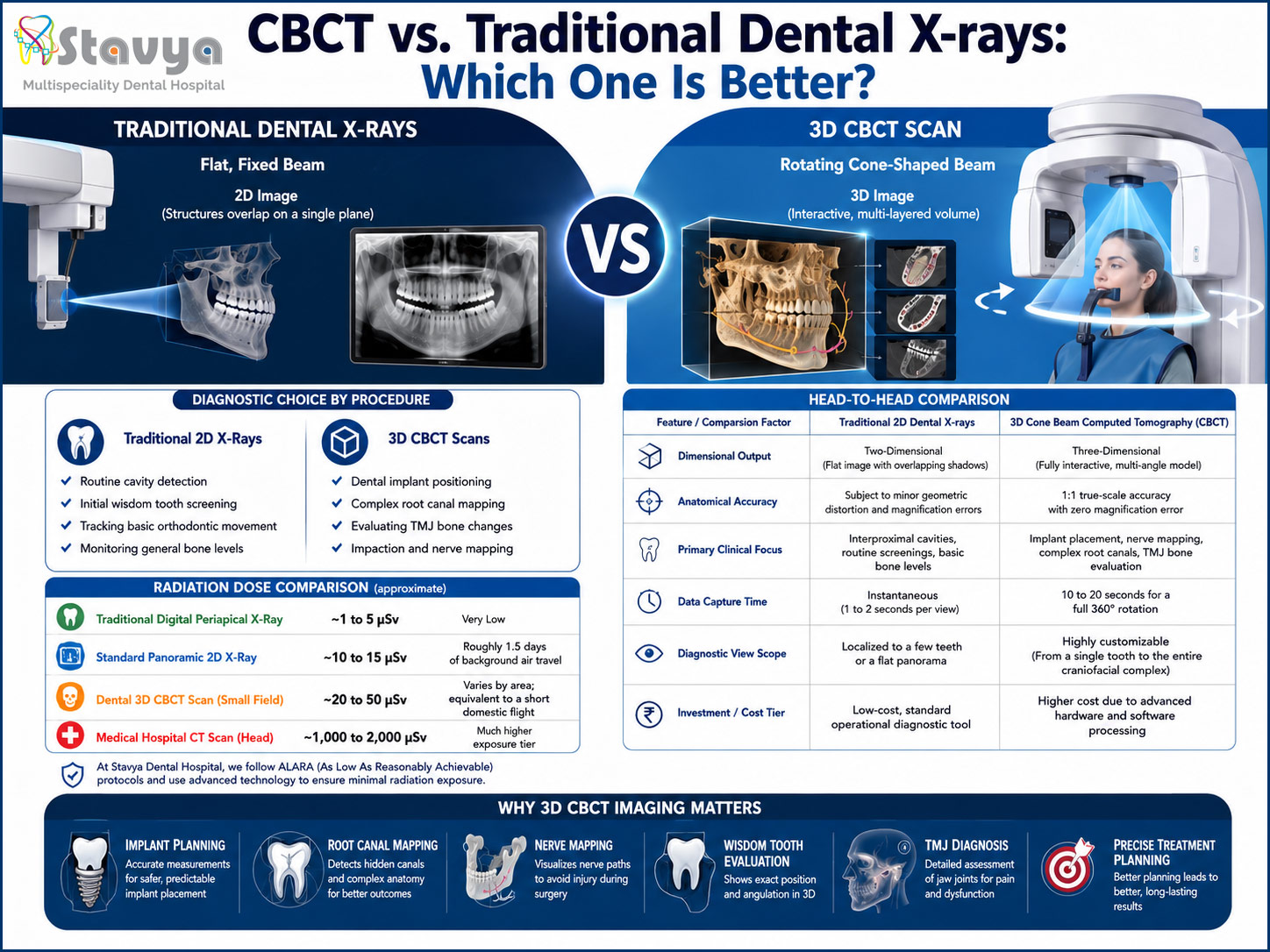
[Traditional X-Ray] -> Flat, Fixed Beam -> 2D Image (Structures overlap on a single plane)

[CBCT Scan]          -> Rotating Cone-Shaped Beam -> 3D Image (Interactive, multi-layered volume)
Traditional 2D Dental X-rays
Traditional digital X-rays—such as bitewing, periapical, or panoramic panoramic (Panorex) views—rely on a flat, fixed radiation beam directed through the oral structures onto a digital sensor or film.
The primary limitation of this method is anatomic superposition. Because a 2D X-ray flattens a three-dimensional object onto a single flat plane, all the structures in the beam’s path overlap. The cheekbones can mask the roots of upper molars, and dense bone can hide early-stage bone loss or infections, requiring dentists to read between the lines of overlapping shadows.
3D Cone Beam Computed Tomography (CBCT)
A CBCT scanner utilizes a completely different approach. Instead of a flat, narrow beam, the machine emits a cone-shaped X-ray beam. During a quick scan—typically lasting between 10 to 20 seconds—the arm of the CBCT machine rotates 360° around the patient’s head.
As it rotates, the scanner captures hundreds of high-resolution, thin data slices (known as voxels). Specialized software compiles these slices to build a highly detailed, 3D digital model of your jaw, teeth, roots, nerve canals, and sinuses. The resulting model can be rotated, sliced, and zoomed into from any angle, allowing clinicians to view your anatomy with 1:1 structural accuracy.
2. Clinical Applications: When Is CBCT Essential?
While traditional X-rays remain highly effective for routine screening, certain complex dental procedures require the exceptional detail that only a 3D CBCT scan can provide.
                 [DIAGNOSTIC CHOICE BY PROCEDURE]
                                |
     +--------------------------+--------------------------+
     |                                                     |
[Traditional 2D X-Rays]                               [3D CBCT Scans]
 * Routine cavity detection                           * Dental implant positioning
 * Initial wisdom tooth screening                     * Complex root canal mapping
 * Tracking basic orthodontic movement                * Evaluating TMJ bone changes
 * Monitoring general bone levels                     * Impaction and nerve mapping
Dental Implant Planning and Surgical Guides
Placing a dental implant is a micro-precise surgical procedure. The titanium post must be embedded into the jawbone at an exact angle and depth to avoid piercing vital anatomical structures, such as the inferior alveolar nerve in the lower jaw or the maxillary sinuses in the upper jaw.
A 2D X-ray can show the height of the bone, but it cannot show its width or density. A CBCT scan allows oral surgeons to measure the exact thickness and quality of your bone in three dimensions. Using this data, specialists can design customized, computer-generated surgical guides, transforming a complex procedure into a highly predictable, minimally invasive surgery.
Endodontics (Complex Root Canal Treatments)
The root canal system inside a tooth is rarely a simple, straight tube. It is often a maze of winding pathways, microscopic lateral branches, and extra hidden canals. Missing a single microscopic canal during treatment can leave bacteria behind, leading to re-infection and root canal failure.
Traditional X-rays often miss extra canals because they hide directly behind larger root structures. A CBCT scan lets the endodontist slice through the tooth digitally, layer by layer, exposing every hidden canal and calculating its exact curvature before treatment ever begins.
Wisdom Tooth Extraction and Nerve Mapping
Lower wisdom teeth frequently develop deep inside the jawbone, often resting directly against or wrapping around the inferior alveolar nerve—the main pathway responsible for providing feeling to your lower lip, chin, and tongue.
Extracting an impacted tooth without knowing its exact 3D relationship to this nerve introduces unnecessary risk. A 2D X-ray shows if the root overlaps the nerve path visually, but it cannot show if the root is sitting in front of, behind, or completely surrounding the nerve. A CBCT scan provides absolute clarity on this relationship, allowing oral surgeons to plan a safe extraction path that protects the nerve from trauma.
Temporomandibular Joint (TMJ) Diagnostics
The temporomandibular joints—the hinge joints connecting your lower jaw to your skull—are complex anatomical structures. Chronic jaw pain, clicking, or popping can stem from microscopic bone erosion, joint misalignment, or disc issues within these structures. CBCT scans provide high-definition, 3D cross-sections of the TMJ bone contours, helping specialists accurately diagnose joint disorders and build effective therapeutic bite splints.
3. Head-to-Head Comparison: CBCT vs. Traditional Dental X-rays
To evaluate which imaging method is best suited for your specific care, it helps to compare them side-by-side across key clinical, safety, and logistical parameters:
| Feature / Comparison Factor | Traditional 2D Dental X-rays | 3D Cone Beam Computed Tomography (CBCT) |
| Dimensional Output | Two-Dimensional (Flat image with overlapping shadows) | Three-Dimensional (Fully interactive, multi-angle model) |
| Anatomical Accuracy | Subject to minor geometric distortion and magnification errors | 1:1 true-scale accuracy with zero magnification error |
| Primary Clinical Focus | Interproximal cavities, routine screenings, basic bone levels | Implant placement, nerve mapping, complex root canals, TMJ bone evaluation |
| Data Capture Time | Instantaneous (1–2 seconds per view) | 10–20 seconds for a full 360° rotation |
| Diagnostic View Scope | Localized to a few teeth or a flat panorama | Highly customizable (From a single tooth to the entire craniofacial complex) |
| Investment / Cost Tier | Low-cost, standard operational diagnostic tool | Higher cost due to advanced hardware and software processing |
4. The Safety Debate: Understanding Radiation Dosage
A common question among patients centers on radiation safety: Does a 3D CBCT scan expose me to significantly more radiation than a traditional X-ray?
To put this in perspective, it helps to understand how medical radiation is measured. The standard unit for measuring radiation absorption in tissue is the Microsievert (µSv). Every human on Earth absorbs a baseline level of natural background radiation daily from the soil, outer space, and building materials, averaging about 8–10 µSv per day.
[Traditional Digital Periapical X-Ray]  --> 1–5 µSv (Very Low)
[Standard Panoramic 2D X-Ray]          --> ~10–15 µSv (Roughly 1.5 days of background air travel)
[Dental 3D CBCT Scan (Small Field)]     --> ~20 to 50 µSv (Varies by area; equivalent to a short domestic flight)
[Medical Hospital CT Scan (Head)]       --> ~1,000 to 2,000 µSv (Much higher exposure tier)
While a CBCT scan does emit slightly more radiation than a single 2D dental film, it is critically important to differentiate a Dental CBCT from a traditional Medical Hospital CT Scan. A hospital CT scan requires massive radiation doses to penetrate dense internal organs and soft tissue layers. A dental CBCT scan, by contrast, utilizes pulse technology and a concentrated beam that targets only the necessary maxillofacial structures, keeping exposure levels incredibly low.
At Stavya Dental Hospital, we strictly follow the ALARA (As Low As Reasonably Achievable) safety protocol. We only prescribe 3D CBCT imaging when the diagnostic information it provides directly improves the safety and success of your treatment. Furthermore, our modern digital machines utilize ultra-fast scanning speeds and customized fields of view to minimize radiation exposure while delivering pristine diagnostic clarity.
5. Which One Is Better? The Ultimate Verdict
So, which technology wins the comparison? The definitive clinical answer is: Neither is universally “better” on its own—they are complementary tools designed for entirely different diagnostic goals.
- Traditional X-rays are the ideal frontline tool for routine preventive care. They are quick, cost-effective, offer incredibly low radiation exposure, and provide all the data a dentist needs to detect early-stage cavities between your teeth or monitor general bone health during your six-month checkup.
- CBCT is the superior tool when accuracy cannot be compromised during specialized treatments. When your care involves placing an implant next to a vital nerve, tracking a hidden root infection, or navigating complex bone anatomy, relying on a flat 2D image leaves too much to chance. In these scenarios, a 3D scan provides the depth and precision needed to ensure a safe, successful outcome.
6. Precision Imaging and Advanced Care at Stavya Dental Hospital
At Stavya Dental Hospital, we believe that exceptional dental treatment begins with an accurate diagnosis. Our facility is equipped with state-of-the-art 3D CBCT digital imaging technology and high-definition intraoral scanners, allowing our multidisciplinary team of specialists to diagnose and plan complex treatments under one roof.
We focus on delivering transparent, patient-guided care. Using our advanced 3D software, our dentists sit down with you to look at your digital model together, guiding you through a step-by-step breakdown of your teeth and bone structures. This interactive approach removes the guesswork from your treatment, helping you make informed decisions for your long-term health.
Schedule Your Advanced Diagnostic Consultation Today
Whether you require a routine preventive checkup or are exploring advanced restorative treatments like dental implants, our specialized team is here to guide you. Contact us today to experience the precision of modern, technology-driven dental care.
- Official Website: www.stavyadental.com
- Direct Clinic Helpline: +91 8980395039