For generations, the dental X-ray has been an indispensable part of oral healthcare. Because more than half of each tooth structure sits hidden beneath the gum line and inside the jawbone, a clinical visual examination can only tell a dentist part of the story. To identify interproximal cavities, map out nerve pathways, evaluate bone density, and track the progress of erupting teeth, dental professionals must rely on diagnostic imaging.
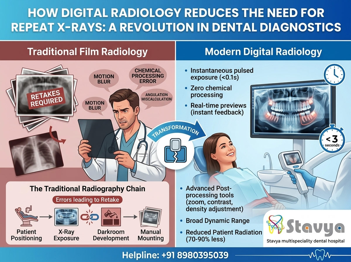
However, traditional film-based radiography long carried a significant operational drawback: the frequent need for repeat X-rays. If a patient moved slightly, if the chemical processing tank was a degree too warm, or if the film was angled incorrectly, the resulting image would turn out blurry, too dark, or distorted. This forced the dental team to restart the process, extending appointment times and subjecting patients to additional radiation exposure.
The introduction of digital radiology has transformed this landscape. By replacing chemical film with high-sensitivity digital sensors and pairing them with advanced imaging software, modern dental diagnostics have drastically cut down the need for retakes.
At Stavya Dental Hospital, our core practice is built on diagnostic precision, clinical efficiency, and patient safety. In this comprehensive, evidence-based guide, we explore the science behind digital radiology, look at why traditional film systems fail, and explain exactly how digital technology reduces repeat X-rays to ensure a safer, more streamlined care experience.
1. The Real Cost of Repeat X-rays in Traditional Dentistry
To understand the value of digital advancement, it helps to examine the mechanics and vulnerabilities of traditional film radiography. Film-based systems rely on a chain of manual, highly sensitive steps where a mistake at any stage can ruin the final image.
                  [THE TRADITIONAL RADIOGRAPHY CHAIN]
                                   |
[Patient Positioning] -> [X-Ray Exposure] -> [Darkroom Development] -> [Manual Mounting]
                                   |
               (Errors at any stage = Invalidation & Retake)
In a traditional setup, several common issues routinely lead to an unusable image:
Processing and Chemical Errors
Traditional X-rays require a physical film packet to be taken into a darkroom or an automated chemical processor. The film must pass through developer, fixer, and water solutions. If the chemical developer is old, contaminated, or mixed at the incorrect temperature, the image will emerge chemically stained, unreadably light, or completely blacked out.
Operator Positioning and Angulation Mistakes
Capturing a precise view of a tooth root requires aligning the X-ray tube head at an exact geometric angle relative to the tooth and the film packet. If the angle is off by just a few degrees, the resulting image suffers from elongation (making the tooth look unnaturally long) or foreshortening (making it look squished). Because these distortions obscure actual anatomical dimensions, the image must be thrown out and retaken.
Patient Movement and Blurring
Traditional dental film has a relatively low sensitivity to radiation, meaning the X-ray machine’s exposure time must be longer to capture a readable image. During this exposure window, if a patient swallows, shifts their jaw, or moves their head slightly, the image suffers from motion blur, rendering it useless for detecting fine details like early-stage decay.
The Hidden Impact on Patients
Every necessary retake adds up. For the patient, repeat X-rays mean:
- Extended time spent in the dental chair.
- Increased frustration and anxiety.
- Accumulated, unnecessary exposure to ionizing radiation.
While a single dental X-ray uses a very low dose of radiation, the biological standard of care—ALARA (As Low As Reasonably Achievable)—mandates that dental teams eliminate unnecessary exposure whenever possible.
2. The Mechanics of Digital Radiology: Why It Changes Everything
Digital radiology removes film and chemicals from the equation entirely. Instead of a plastic packet containing radiation-sensitive film, the dentist places a slim, highly advanced electronic digital sensor (using Charge-Coupled Device [CCD] or Complementary Metal-Oxide-Semiconductor [CMOS] technology) or a Photostimulable Phosphor (PSP) plate inside your mouth.
[Digital Sensor Placement] -> [Instantaneous Pulsed Exposure] -> [Immediate On-Screen Display]
When the X-ray beam meets the digital sensor, the sensor’s electronic matrix converts the radiation energy into an instant electronic signal. This signal is sent directly to an attached computer system, rendering a high-definition, crisp image on the monitor within seconds.
By bypassing the darkroom and utilizing hyper-sensitive electronic receptors, digital radiology eliminates the vulnerabilities that make traditional X-rays prone to failure.
3. How Digital Radiology Explicitly Eliminates Repeat X-rays
Digital technology directly tackles the root causes of film errors, dropping the rate of repeat X-rays close to zero through several key engineering and operational features:
1. Complete Elimination of Chemical Processing
Because digital radiology relies entirely on electronic data transfer, the darkroom, developer fluids, fixer solutions, and manual drying racks are completely obsolete. By removing this complex chemical chain, 100% of processing-related errors are permanently eliminated. There are no expired chemicals to blur an image, no darkroom light leaks to ruin a film, and no chemical stains to obscure a diagnosis.
2. Real-Time Image Previews for Instant Verification
With traditional film, a dentist or hygienist has to wait 5 to 8 minutes for the film to develop before discovering that a patient shifted or an angle was slightly off. If an error occurred, the entire setup had to be rebuilt from scratch.
Digital systems display the captured image on a high-resolution chairside screen in less than 3 seconds. This instant feedback allows the clinician to verify proper positioning, coverage, and clarity immediately. If a minor correction is needed, it can be handled instantly without disrupting the flow of your appointment.
3. Hyper-Wide Dynamic Range (Exposure Latitude)
Traditional film requires a very specific, narrow amount of radiation to create a clear image. If the exposure time is a fraction of a second too short, the film is severely underdeveloped (too light); if it is a fraction too long, it is overexposed (too dark).
Digital sensors possess an incredibly broad dynamic range. They can capture accurate details even if the exposure settings aren’t perfectly dialed in. The software automatically compensates for minor exposure variances, processing the raw data into a clean, balanced image and saving the patient from a retake.
4. Advanced Post-Processing Software Tools
When a traditional film comes out slightly too light or lacks contrast, it cannot be altered—the patient must undergo another X-ray.
Digital radiology software gives clinicians a powerful toolkit to adjust and enhance images after they are taken:
- Contrast Enhancement: Easily separate dense enamel from softer tooth layers to spot early cavities.
- Magnification and Zoom: Zoom in on a single root tip to check for deep infections without needing a separate, localized view.
- Colorization and Inversion: Toggle between positive and negative views to evaluate bone density changes around dental implants.
- Density Adjustment: Lighten or darken specific areas of the image dynamically on screen to reveal hidden structural details.
Because the software can rescue and optimize a suboptimal image, the need to take a fresh X-ray simply to see a detail better is completely eliminated.
5. Ultra-Short Exposure Times
Digital sensors are substantially more sensitive to radiation than traditional dental film. Because they require far less energy to create a high-definition image, the X-ray machine’s exposure time can be cut by 70% to 90%.
Instead of an exposure lasting a full second or more, a digital pulse takes just a tiny fraction of a second. This incredibly fast capture time virtually eliminates motion blur caused by accidental patient movement, making it an ideal solution for young children, elderly individuals, or patients dealing with dental anxiety.
4. Side-by-Side Comparison: Digital vs. Traditional Film Radiology
To see how these two diagnostic approaches compare across key performance, safety, and workflow metrics, review this detailed breakdown:
| Diagnostic Feature / Consideration | Traditional Film Radiology | Modern Digital Radiology |
| Image Capture Speed | 5 to 8 minutes (Requires chemical processing) | Instantaneous (Renders on-screen in < 3 seconds) |
| Primary Source of Retakes | Chemical errors, darkroom leaks, minor movement | Exceptionally rare (Limited to severe positioning errors) |
| Radiation Exposure Profile | Baseline standard exposure | Reduced by 70% to 90% compared to film |
| Image Modification Capabilities | None; image is permanently fixed on plastic film | Extensive (Real-time zoom, contrast adjustments, sharpening) |
| Storage & Record Security | Physical jackets (Prone to loss, fading, or damage) | Encrypted digital files (Instantly accessible and safely backed up) |
| Environmental Footprint | High (Involves toxic lead foil and chemical waste disposal) | Eco-Friendly (Zero chemical waste or heavy metal disposal) |
5. Beyond Reducing Retakes: Added Benefits of Digital Radiology
While cutting down the need for repeat X-rays is a massive win for safety and efficiency, digital imaging offers several additional advantages that elevate the standard of modern dental care:
Enhanced Patient Education and Visual Clarity
Traditional dental films are tiny, roughly the size of a postage stamp. When a dentist holds a small film up to a lightbox and points out a microscopic cavity, it can be incredibly difficult for a patient to see or comprehend the issue.
Digital X-rays are projected onto large, high-definition chairside monitors. Your dentist can zoom in, highlight specific areas, and show you exactly what is happening inside your tooth in an easy-to-understand format. This visual clarity empowers you to make informed decisions about your treatment plan with complete confidence.
Streamlined Specialist Collaborations
If your dental care requires input from a specialist—such as an oral surgeon, periodontist, or endodontist—traditional film requires physically mailing the records or asking the patient to transport them by hand. This process wastes time and risks losing vital diagnostic data. Digital X-rays are saved as secure, encrypted image files that can be shared instantly with trusted medical specialists, ensuring seamless, coordinated care.
A Greener, More Sustainable Practice
Traditional radiography requires using toxic developer fluids, chemical fixers, and protective lead foils, all of which generate hazardous waste that must be carefully managed to protect local ecosystems. Digital radiology is entirely eco-friendly, operating with zero hazardous chemicals or physical waste.
6. Comprehensive Diagnostic Care at Stavya Dental Hospital
At Stavya Dental Hospital, we treat diagnostic precision as a fundamental pillar of long-term oral health and patient safety. Our clinical facility is equipped with advanced, low-radiation digital radiology systems, high-definition intraoral sensors, and advanced 3D CBCT imaging platforms.
By removing the guesswork, chemical delays, and inefficiencies of traditional film systems, our experienced team provides a streamlined, comfortable, and highly precise diagnostic experience. We design your care path using real-time, high-definition imaging, ensuring that your treatment is built on an exceptionally accurate understanding of your smile.
Experience Safe, Technology-Driven Dental Care
Ready to experience the precision, safety, and comfort of modern digital dental diagnostics? Schedule your comprehensive preventative dental evaluation with our specialist team today.
- Official Website: www.stavyadental.com
- Direct Clinic Helpline: +91 8980395039